

“Shoulder mechanics” is often used as if it were one measurement. It is not. The phrase can refer to bone position, joint motion, external force, net joint moment, model-estimated reaction force, muscle activity, symptoms or injury. Those outcomes are related, but they are not interchangeable.
Surface design enters this record at the posterior torso. The bench can change contact, clearance, effective height, resistance to sliding and the setup available to the athlete. That makes it a legitimate mechanical variable. It does not let geometry alone prove that a surface is safer, therapeutic or universally superior.
Layer 01 · Anatomy
How the Shoulder’s Parts Work Together
The humeral head articulates with the glenoid at the glenohumeral joint. The scapula moves relative to the thorax and connects through the acromioclavicular joint to the clavicle; the clavicle connects to the sternum at the sternoclavicular joint. Rotator-cuff muscles contribute direction-specific control, while larger muscles generate and transmit pressing force.
Context: why a visible scapular pattern is not a diagnosis
Scapular position and motion are clinically relevant, but reviews and consensus statements also warn against treating one visible scapular pattern as a diagnosis or a universal mechanism.[1][2] Bench-press interpretation therefore has to specify the movement, load, athlete and measurement method.
AMM Research · Anatomical orientation
The shoulder chain and its support boundary
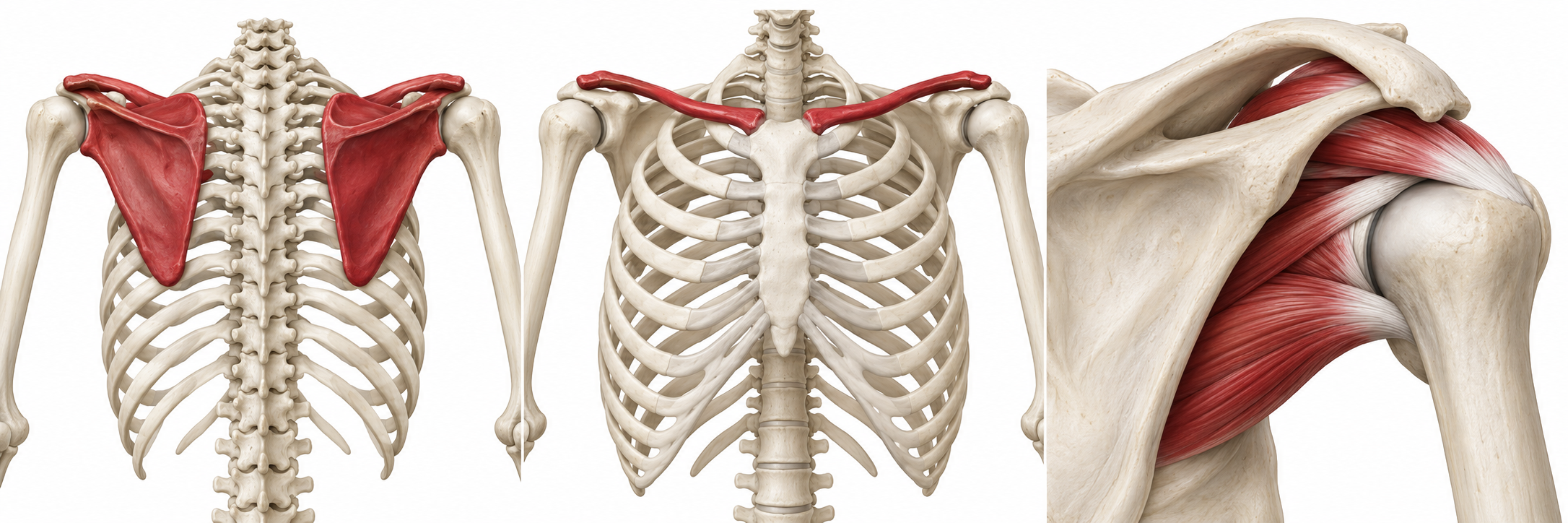
Posterior view
Scapula on the thorax
Both shoulder blades lie on the posterior rib cage. The socket-bearing scapula connects the upper arm to the shoulder girdle.
Anterior view
Clavicle, SC and AC joints
The sternoclavicular (SC) joint connects the clavicle to the sternum. The acromioclavicular (AC) joint links its lateral end to the scapula.
Shoulder detail
Glenoid, humerus and cuff
The humeral head meets the glenoid. Cuff muscles and tendons contribute to control around this articulation.
The bench meets the back of the torso. Contact and edge clearance belong to the interface record.
See the equipment contact example →Force at the hands contributes to the external loading record. Its magnitude needs a defined measurement.
Force along the bar can alter calculated moments even when the visible path looks similar.
Follow the force record →Interpretation: the surface is one boundary in a linked system. It may alter contact or available pose; motion, force, muscle activity and symptoms still need their own measurements.
Layer 02 · Kinematics
First Ask What Moved
Three distinct records
Name the motion before drawing a conclusion
Bar
Path, displacement and timing.
Shoulder girdle
Scapular translation, rotation and tilt.
Upper arm / torso
Humeral excursion and thoracic posture.
Kinematics describes position and motion: humeral elevation and rotation, scapular rotation and tilt, thoracic posture, bar path and joint excursion. A bench surface may change the pose an athlete can establish or the contact encountered as the upper arm moves. A camera view or visible edge clearance can generate that question, but motion capture or imaging must answer it.
Motion capture · imaging · video-based kinematics
Angles · translations · paths · timing
Movement is not a direct measurement of joint force, tissue stress, pain or injury.
What “scapular freedom” needs to specify
“Scapular freedom” is therefore too broad unless it is operationalized. Retraction, protraction, upward rotation, internal or external rotation, tilt, translation and clearance are different phenomena. More motion is not automatically better; pressing also requires task-specific control.
Layer 03 · Kinetics and models
Then Ask What Force or Moment Was Estimated
Instrumented bar · inverse dynamics · musculoskeletal modeling
External forces · net joint moments · model-estimated reaction forces and muscle demand
Model outputs depend on assumptions; they are not in-vivo tissue measurements or clinical outcomes.
Read the participant and measurement protocol
Noteboom and colleagues tested 10 healthy, experienced strength athletes—nine men and one woman, with 6.7 ± 3.9 years of bench experience—across 21 combinations. Grip widths were 1.0, 1.5 and 2.0 times bi-acromial width; shoulder-abduction targets were 45°, 70° and 90°; scapular conditions were neutral, retracted and released. Participants completed three repetitions with a 16 kg bar. Motion capture and an instrumented bar supplied inputs to an OpenSim shoulder model.[3]
Noteboom et al. · 2024 · Table 2
Technique changes the modeled demand
- 10 experienced lifters
- 21 technique combinations
- 16 kg bar
- 3 repetitions per trial
One selected modeled muscle part, compared across three technique components. Every panel uses the same 0–1 scale; BAW means bi-acromial width.
Grip width
SD 0.20 relative activity
SD 0.17 relative activity
SD 0.13 relative activity
relative activity · bars show means; SD printed for each condition
Scapular pose
SD 0.17 relative activity
SD 0.17 relative activity
SD 0.16 relative activity
relative activity · bars show means; SD printed for each condition
Abduction target
SD 0.17 relative activity
SD 0.17 relative activity
SD 0.16 relative activity
relative activity · bars show means; SD printed for each condition
Body and bar kinematics; vertical and mediolateral bar forces.
Selected glenohumeral and acromioclavicular reaction forces; selected muscle activity.
In-vivo joint force, ligament loading, pain, injury, production bench-pad differences or heavy working sets.
Read the joint-force findings and model limitations
Narrower grips and scapular retraction reduced selected acromioclavicular compression, glenohumeral posterior shear and modeled rotator-cuff activity during parts of the repetition. Mediolateral bar force materially affected the estimates. The released condition used a pool noodle under the spine, and released versus neutral conditions did not differ in modeled joint-reaction force. Twelve percent of trials were excluded for missing marker or lateral-force data, and ligaments were absent from the model.[3]
The correct conclusion is specific: technique changed selected model-estimated shoulder loads. The study did not test a production support surface or calculate an individual injury probability.
The Visible Bar Path Is Not the Whole Force Record
Force direction · measurement framework
A bar path does not contain the whole force record
Record the external forces
Vertical and mediolateral force components at the bar.
Combine with body geometry
Joint positions and moment arms through the repetition.
Interpret the calculated outcome
Net joint moments or model-estimated reaction forces, under stated assumptions.
In 35 strength-trained adults, Mausehund and colleagues found condition differences of up to 12% in load, up to 43% in shoulder net moments with wider grips and up to 26% in elbow moments as grip narrowed. Their central methodological point was that lateral bar forces materially change calculated joint moments.[4]
Related evidence: grip strategy and torso technique
Larsen and colleagues confirmed that grip affected both the weight lifted and the horizontal-force strategy during 1RM pressing in 14 experienced men.[5] Cudlip and colleagues added a torso-technique comparison: in 20 experienced men at 25%, 50% and 75% of self-reported 1RM, the powerlifting arch increased latissimus dorsi activity, while the standardized technique produced approximately 8% larger integrated shoulder moments.[6] An arch does not make shoulder demand simply “more” or “less”; it redistributes a multi-joint task.
Layer 04 · Muscle activity
Electrical Activity Is Direction-Specific
Anatomy context · Muscle roles
Several muscles contribute to the press
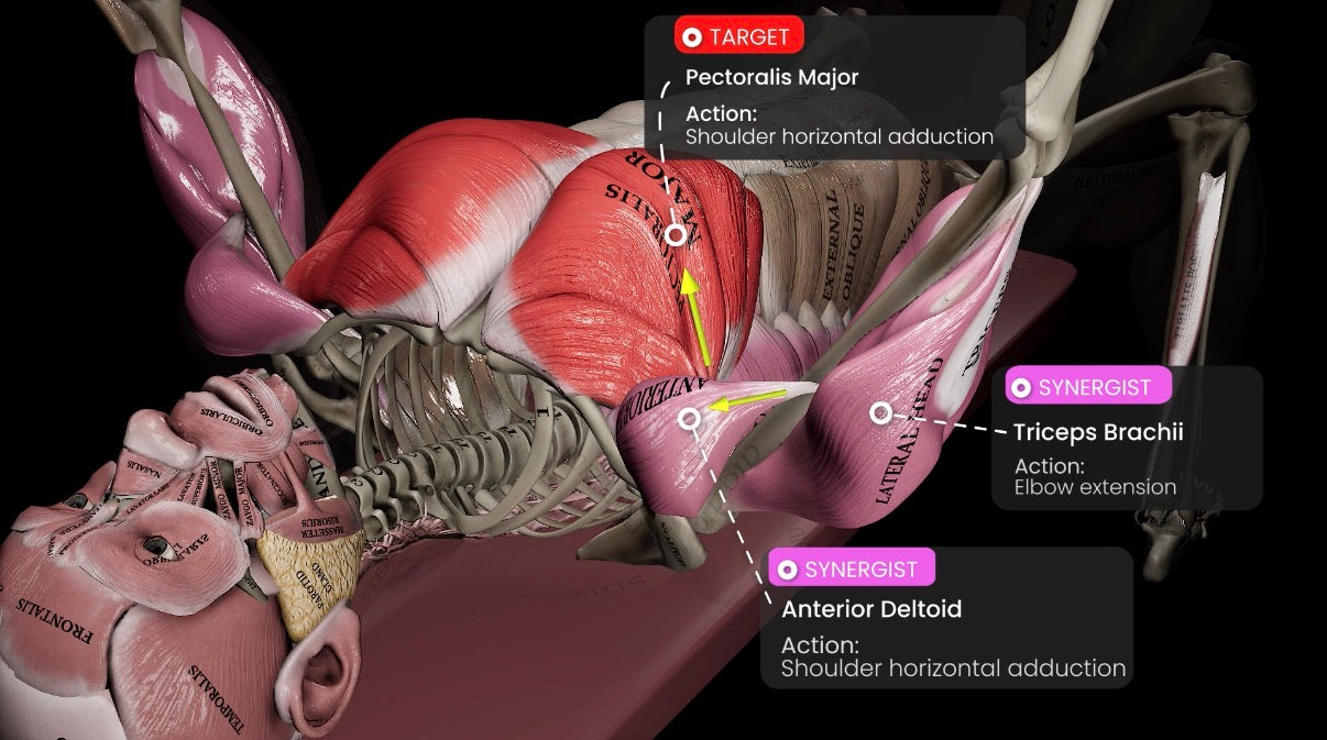
Moves the upper arm
Helps bring the upper arm across the chest during the press.
Assists shoulder motion
Contributes to shoulder motion; its role depends on the task and position.
Extends the elbow
Contributes to straightening the elbow during the upward press.
Surface or fine-wire electromyography
Recorded electrical amplitude and timing for sampled muscles
EMG is not direct joint force, hypertrophy, pain or injury risk.
Wattanaprakornkul et al. · 2011 · EMG comparison
Pressing and rowing recruit the cuff differently
Bench press
Infraspinatus activity exceeded subscapularis activity.
Row
Subscapularis activity exceeded infraspinatus activity.
Read the muscle-recording protocol and full explanation
Wattanaprakornkul and colleagues recorded nine shoulder muscle sites in 15 participants during bench press and row tasks at 20%, 50% and 70% of maximum load. Infraspinatus activity exceeded subscapularis activity during the bench press, while the pattern reversed during the row.[7] The cuff response was reciprocal and task-direction-specific—not one generic “stability” signal.
The study informs shoulder control during pressing but did not compare bench surfaces or track symptoms. A higher or lower electrical signal, by itself, does not identify the better exercise or safer joint.
Layer 05 · Surface interaction
Where Contact With the Bench Enters the Mechanics
A bench establishes contact width, contour, friction, compliance, effective height and an edge around the posterior torso. These properties can affect the setup an athlete uses, resistance to sliding, rack relationship and the contact encountered near the scapular region. They are legitimate independent variables.
AMM Research · Visual 25Equipment photograph
A photograph locates contact; instruments measure it
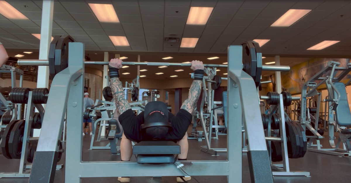
The upper back is supported while the hands connect to the bar. The photograph cannot reveal internal joint forces, quantify shoulder-blade movement or establish a safety outcome.
Advanced Muscle Mechanics product-image archive. Original image, unaltered.
Where and how much of the thorax and pelvis are supported under load?
Which edge or contour approaches the upper torso through the repetition?
How do friction and compression affect position or drift?
Do measured kinematics, force, EMG or performance change?
Surface geometry can answer the first two questions descriptively. It cannot answer the fourth by inspection. The Athlete-to-Bench Interface owns the complete direct evidence progression; here, only the shoulder-relevant acute record is interpreted.
Direct product evidence · interpreted at the measured layer
The Acute Launch Pad Study Changed the Support Condition
In the randomized crossover by Kidwell and colleagues, 10 resistance-trained men performed five repetitions at 70% of each participant’s established 1RM on a conventional flat bench and with the Launch Pad.[8] The Launch Pad condition produced higher bilateral pectoralis sEMG, mean and peak bar velocity and vertical displacement. Concentric power was 249 ± 103 W versus 271 ± 93 W, but the difference was not statistically significant (p = .071).
AMM Research · Measurement and meaning
Keep the measurement attached to its meaning
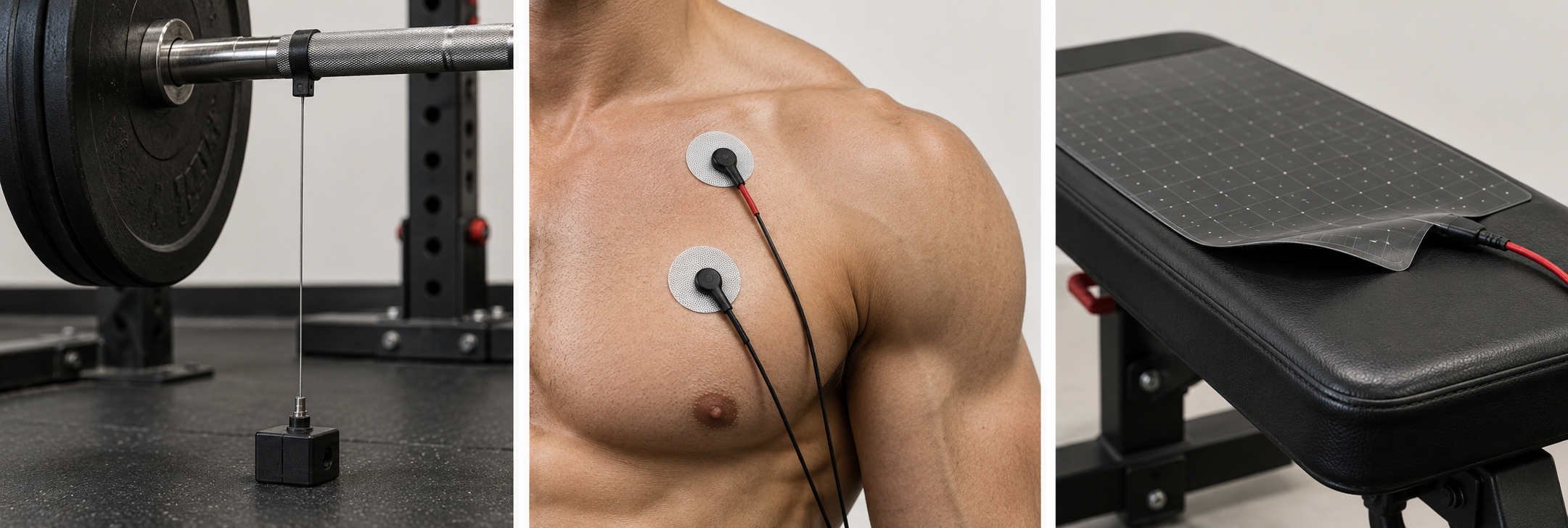
01 · Observation
Bar moves
- Measurement
- Position + time
- Interpretation
- Bar kinematics
02 · Observation
Muscle is active
- Measurement
- Surface EMG
- Interpretation
- Electrical activity
03 · Observation
Support changes
- Measurement
- Contact / pressure
- Interpretation
- Interface condition
Kidwell et al. · 2026 · Table 1
Six measured outcomes, with the full result in view
- 10 trained men
- 70% 1RM
- 5 repetitions
- Randomized crossover
Right pectoralis sEMG
SD 24.8 % MVC
SD 20.1 % MVC
% MVC · bars show means; SD printed for each condition
Left pectoralis sEMG
SD 23.4 % MVC
SD 20.0 % MVC
% MVC · bars show means; SD printed for each condition
Mean bar velocity
SD 0.09 m/s
SD 0.09 m/s
m/s · bars show means; SD printed for each condition
Peak bar velocity
SD 0.11 m/s
SD 0.09 m/s
m/s · bars show means; SD printed for each condition
Vertical bar displacement
SD 7 cm
SD 4 cm
cm · bars show means; SD printed for each condition
Concentric power
SD 103 W
SD 93 W
W · bars show means; SD printed for each condition
View the original compact numerical summary
Pectoralis sEMG · bar velocity · vertical displacement · concentric power
Scapular kinematics · shoulder-joint reaction force · rotator-cuff EMG · pain · injury
The study establishes that this specific support intervention was not mechanically neutral for the measured repetition. It does not show how the scapula moved, how joint loading changed or whether the condition prevents or treats shoulder problems.
Layer 06 · Clinical meaning
What the Total Shoulder Record Allows Us to Say
Grip, force direction, scapular pose, arch and a tested support condition can change selected mechanical or EMG outcomes under defined protocols.
Surface contact and clearance may interact with shoulder pose, motion and force strategy. That relationship needs direct kinematic and kinetic measurement.
No cited surface study shows injury prevention, pain treatment or a universally safer geometry.
Pain, function, time loss, injury incidence and return to sport require clinical outcomes and appropriate follow-up.
Why mechanical findings cannot substitute for clinical outcomes
This distinction matters because biomechanical language is often converted too quickly into clinical language. A reduction in a modeled force component is not equivalent to lower injury incidence. A larger EMG signal is not equivalent to tissue protection. Greater clearance is not equivalent to symptom relief. Each can justify the next study, but each must stop at the endpoint actually measured.
How individual context limits the group result
Clinical meaning also depends on the person. Group averages from healthy, trained participants cannot determine an individual athlete’s diagnosis, tissue tolerance or return-to-sport readiness. A support condition that is tolerable and repeatable for one athlete may be inappropriate for another; the reason must be evaluated rather than inferred from the product geometry.
For coaching, change one variable when possible and record the condition precisely. For research, match the conclusion to the method. For persistent pain, neurological symptoms, postoperative restrictions or return-to-training decisions, use appropriate clinical assessment rather than a mechanical inference.
Continue the mechanics sequenceScapular Motion During the Bench Press explores the motion terms in greater depthReferences
- Ludewig PM, Reynolds JF. (2009). The association of scapular kinematics and glenohumeral joint pathologies. Journal of Orthopaedic & Sports Physical Therapy, 39(2), 90–104. doi:10.2519/jospt.2009.2808. ↩
- Kibler WB, Ludewig PM, McClure PW, Michener LA, Bak K, Sciascia AD. (2013). Clinical implications of scapular dyskinesis in shoulder injury: the 2013 consensus statement from the Scapular Summit. British Journal of Sports Medicine, 47, 877–885. doi:10.1136/bjsports-2013-092425. ↩
- Noteboom L, Belli I, Hoozemans MJM, Seth A, Veeger HEJ, van der Helm FCT. (2024). Effects of bench press technique variations on musculoskeletal shoulder loads and potential injury risk. Frontiers in Physiology, 15, 1393235. doi:10.3389/fphys.2024.1393235. ↩
- Mausehund L, Werkhausen A, Bartsch J, Krosshaug T. (2022). Understanding bench press biomechanics—The necessity of measuring lateral barbell forces. Journal of Strength and Conditioning Research, 36(10), 2685–2695. doi:10.1519/JSC.0000000000003948. ↩
- Larsen S, Gomo O, van den Tillaar R. (2021). A biomechanical analysis of wide, medium, and narrow grip width effects on kinematics, horizontal kinetics, and muscle activity on the sticking region in recreationally trained males during 1-RM bench pressing. Frontiers in Sports and Active Living, 2, 637066. doi:10.3389/fspor.2020.637066. ↩
- Cudlip AC, Maciukiewicz JM, Pinto BL, Dickerson CR. (2022). Upper extremity muscle activity and joint loading changes between the standard and powerlifting bench press techniques. Journal of Sports Sciences, 40(9), 1055–1063. doi:10.1080/02640414.2022.2046937. ↩
- Wattanaprakornkul D, Halaki M, Cathers I, Ginn KA. (2011). Direction-specific recruitment of rotator cuff muscles during bench press and row. Journal of Electromyography and Kinesiology, 21(6), 1041–1049. doi:10.1016/j.jelekin.2011.09.002. ↩
- Kidwell JA, Yamamoto T, Hetherton KJ, et al. (2026). Acute effects of thoracic-spinal elevation via a novel bench press pad on sEMG and barbell kinetics in resistance-trained males. International Journal of Exercise Science, 19(1), 1003. doi:10.70252/IJES2026103. ↩