

The scapula does not simply retract or “stay down.” It translates around the thorax, rotates in three dimensions and changes its relationship to the clavicle and humerus. During a bench press, muscular force, bar load and reaction from the support surface act on that linked system. The central question is therefore: which scapular variables actually change during a loaded repetition, and which are only assumed from a setup cue?
The focus here is motion vocabulary, bench-phase questions and translation from research to cueing. The linked shoulder-mechanics record covers the deeper anatomy, force-model and clinical-inference boundaries. Keeping those jobs separate prevents a familiar error: treating a visible setup, a model output and an injury claim as if they were the same evidence.
Layer 01 · Name the motion
The Shoulder Blade Moves in Several Directions
Three-dimensional scapular descriptions conventionally include upward/downward rotation, internal/external rotation and anterior/posterior tilt, alongside translations such as protraction/retraction and elevation/depression. Bone-pin work during dynamic shoulder movement helped establish why surface observation alone can miss or distort these motions.[1] That study was not a bench-press experiment; it is an adjacent measurement foundation.
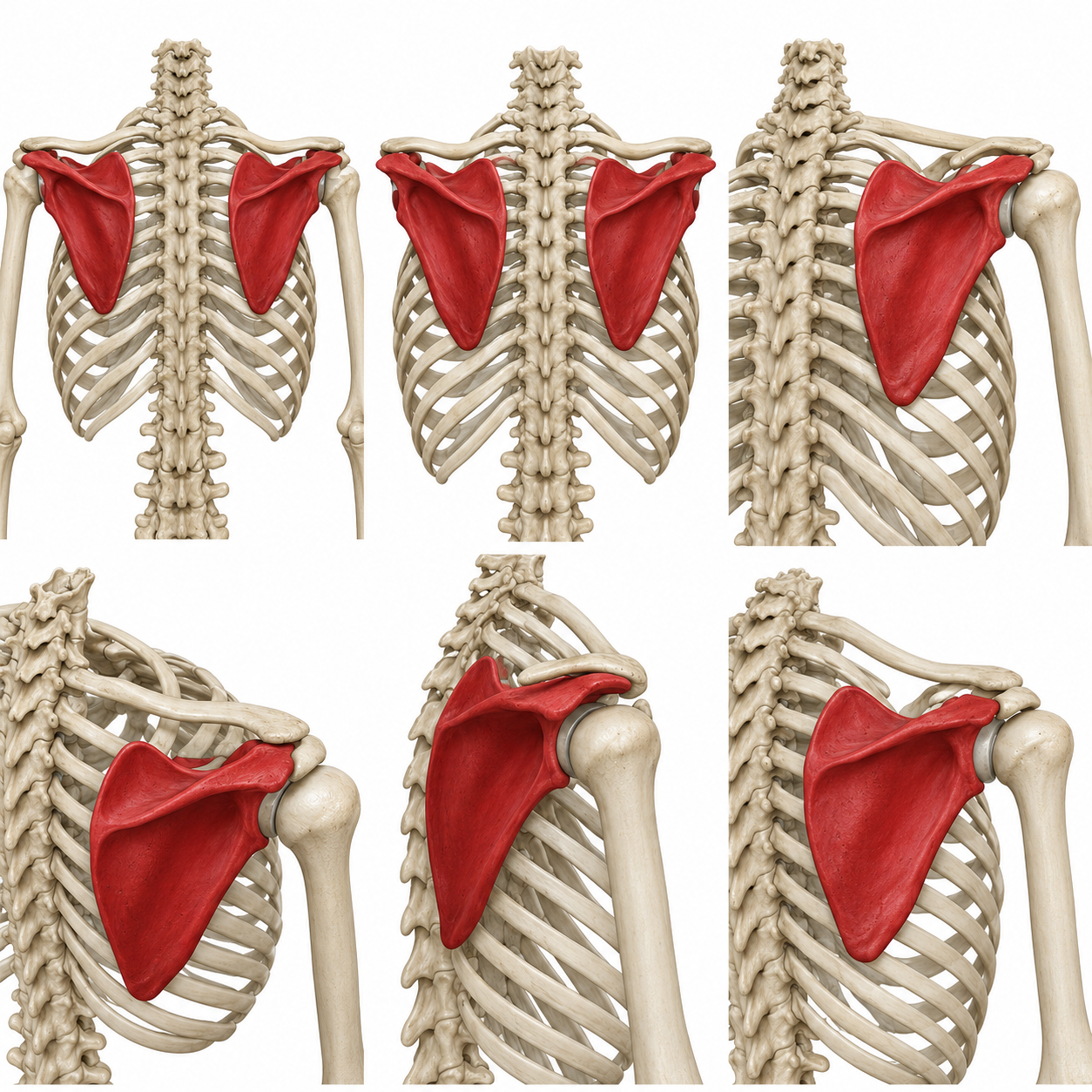
Retraction ↔ protraction
Medial or lateral movement around the thorax—not a complete description of rotation or tilt.
Elevation ↔ depression
Superior or inferior movement; a “down” cue does not guarantee a fixed position under load.
Upward ↔ downward
Changes glenoid orientation in the frontal plane; it is not synonymous with protraction.
Internal ↔ external
Describes orientation relative to the thorax; it requires a defined coordinate system.
Anterior ↔ posterior tilt
Top-to-bottom tipping. A posterior support surface may affect contact without proving a tilt change.
Scapula + clavicle + humerus
Observed bar travel does not reveal how motion was distributed among the linked segments.
These terms are coordinate-dependent and not perfectly interchangeable across methods. Clinical reviews also caution that scapular differences can be associated with shoulder conditions without establishing a single causal pattern.[2] The 2013 Scapular Summit likewise framed dyskinesis as an impairment finding that requires context, not a stand-alone diagnosis.[3]
Layer 02 · Locate the phase
Consider Each Phase of the Lift
A setup cue is often judged by the repetition that follows, yet scapular behavior may differ during the unrack, descent, reversal, ascent and rack. A study that records only barbell outcomes cannot tell whether a scapular pose was maintained. A technique score cannot supply three-dimensional angles. A static setup image cannot reveal phase timing.
Initial torso contact, scapular pose and the perturbation created when the bar leaves the hooks.
Posterior contact rises while humeral position and bar path change.
Peak descent depth and transition timing; a static cue may be most challenged here.
Force direction and humeral motion evolve through the sticking region.
Scapular and torso positions may change again as the athlete reaches the uprights.
The field needs phase-resolved scapular angles, bar forces and surface-contact data collected together. Until then, “the scapula moved more” is incomplete unless the direction, phase, load, method and reference frame are stated.
Measurement method changes the answer
Method detail: how scapular motion is measured
Skin-mounted markers are practical for loaded movement, but the scapula lies beneath layers of moving soft tissue. Acromion marker-set approaches can estimate orientation while the marker set remains secure; palpation-based methods can identify landmarks at selected poses; bone pins and imaging reduce some soft-tissue uncertainty but are invasive, constrained or difficult to combine with heavy lifting. A study’s method therefore determines both the available motion record and the confidence appropriate to it.
Heavy bench research also creates a practical conflict: the bench can hide posterior markers exactly where contact is scientifically interesting. Any future protocol must report missing-data rules, calibration, coordinate systems and whether contact itself compromised the measurement.
Layer 03 · Audit the bench-specific record
What Bench-Specific Studies Show
Retracted versus freer setup
Piepoli and colleagues studied 28 recreationally trained men in randomized-order sessions. They compared a flat/free-scapula condition with a retracted setup across maximal velocity tests at 40%, 60% and 80% 1RM, 1RM testing and repetitions to failure at 70% 1RM.[4]
Piepoli et al. · 2023 · setup comparison
A change in bar travel, with a distinct performance profile
- Upward bar displacement
- Lower with retractionSubmaximal-load comparisons · p ≤ .024
- Mean bar velocity
- No detected condition differencep ≥ .473
- 1RM / repetitions to failure
- No detected condition differencep = .147 / p = .447
Read the complete performance findings
- Retraction produced lower upward bar displacement at submaximal loads (p ≤ .024).
- No condition differences were detected for mean velocity (p ≥ .473), 1RM (p = .147), repetitions to failure (p = .447), RPE or discomfort.
- Performance tended to favor the variant with which each participant was more familiar.
Boundary: the study compared instructed setups and performance outcomes; it did not report three-dimensional scapular kinematics.
Pose, grip and abduction
Noteboom and colleagues tested 10 healthy, experienced lifters through 21 combinations of grip width, shoulder-abduction target and scapular condition with a 16 kg instrumented bar, motion capture and an OpenSim shoulder model.[5]
Noteboom et al. · 2024 · modeled muscle activity
Scapular pose changed the model’s muscular demand
Four selected rotator-cuff parts, with the complete eight-part table below. Every panel uses the same 0–1 scale.
Supraspinatus anterior
SD 0.17 relative activity
SD 0.17 relative activity
SD 0.16 relative activity
relative activity · mean with SD · zero-origin scale
Supraspinatus posterior
SD 0.07 relative activity
SD 0.06 relative activity
SD 0.07 relative activity
relative activity · mean with SD · zero-origin scale
Infraspinatus inferior
SD 0.10 relative activity
SD 0.11 relative activity
SD 0.10 relative activity
relative activity · mean with SD · zero-origin scale
Subscapularis medialis
SD 0.14 relative activity
SD 0.11 relative activity
SD 0.15 relative activity
relative activity · mean with SD · zero-origin scale
View all eight modeled rotator-cuff parts
| Modeled muscle part | Neutral | Retracted | Released |
|---|---|---|---|
| Infraspinatus inferior | 0.28 ± 0.10 | 0.24 ± 0.11 | 0.28 ± 0.10 |
| Infraspinatus superior | 0.33 ± 0.10 | 0.29 ± 0.11 | 0.30 ± 0.10 |
| Teres minor | 0.16 ± 0.06 | 0.14 ± 0.08 | 0.16 ± 0.06 |
| Subscapularis superior | 0.30 ± 0.14 | 0.25 ± 0.13 | 0.30 ± 0.15 |
| Subscapularis medialis | 0.28 ± 0.14 | 0.21 ± 0.11 | 0.27 ± 0.15 |
| Subscapularis inferior | 0.34 ± 0.13 | 0.29 ± 0.14 | 0.32 ± 0.14 |
| Supraspinatus posterior | 0.25 ± 0.07 | 0.19 ± 0.06 | 0.24 ± 0.07 |
| Supraspinatus anterior | 0.62 ± 0.17 | 0.50 ± 0.17 | 0.60 ± 0.16 |
Read the joint-force findings and model context
- Retracted versus neutral conditions reduced selected modeled glenohumeral and acromioclavicular reaction-force components.
- Released versus neutral conditions did not differ in modeled joint-reaction force.
- Outputs depended on the model, force inputs and light-load protocol.
Boundary: this is evidence about selected model estimates—not in-vivo tissue load, symptoms, injury or production pad width.
Montenegro et al. · 2026 · motor learning
The exercise order changed; the training volume was matched
High contextual interference
- Bench set
- Seal rows during the 3-minute rest
- Next bench set
Low contextual interference
- Bench sets and rests
- Finish the bench work
- Seal-row work
Study detail: technique scores, strength and scapular retraction
A 2026 coaching study adds a motor-learning signal rather than a kinematic one. Montenegro and colleagues randomized 15 powerlifters to six weeks of the same volume-matched seal-row and bench training. The high-contextual-interference group performed seal rows during the three-minute rests between bench sets; the low-interference group performed them after the bench work. Both groups improved strength without a group-by-time strength effect. Group-by-time effects were reported for the global bench-technique score (p = .038) and its scapular adduction/retraction item (p = .009), with the high-interference group improving that item at the final test.[6] The item came from a 13-point video-scored technique checklist. It was not a seal-row score, a measured scapular angle or evidence that the pad changed motion.
Scapular setup instructions can alter bar displacement and selected model estimates under defined protocols. Familiarity and task context matter.
No cited study maps heavy bench-press scapular motion across every phase or proves that one scapular cue prevents injury.
Layer 04 · Translate without overclaiming
A Coaching Cue Does Not Measure Movement
Practitioner coaching makes the field problem concrete. In the EFS Bench Manual, Dave Tate organizes the feet, upper back, lats, shoulder blades and pressure on the traps into a stable pressing base that must be practiced and maintained.[7] That is a coaching solution to setup repeatability. It does not establish that the scapula is immobile, identify its three-dimensional orientation or prove that one cue fits every athlete.
AMM Research · From cue to measurement
Keep the instruction and the movement record connected
A matched posterior view makes the shoulder blades easier to locate against the rib cage.
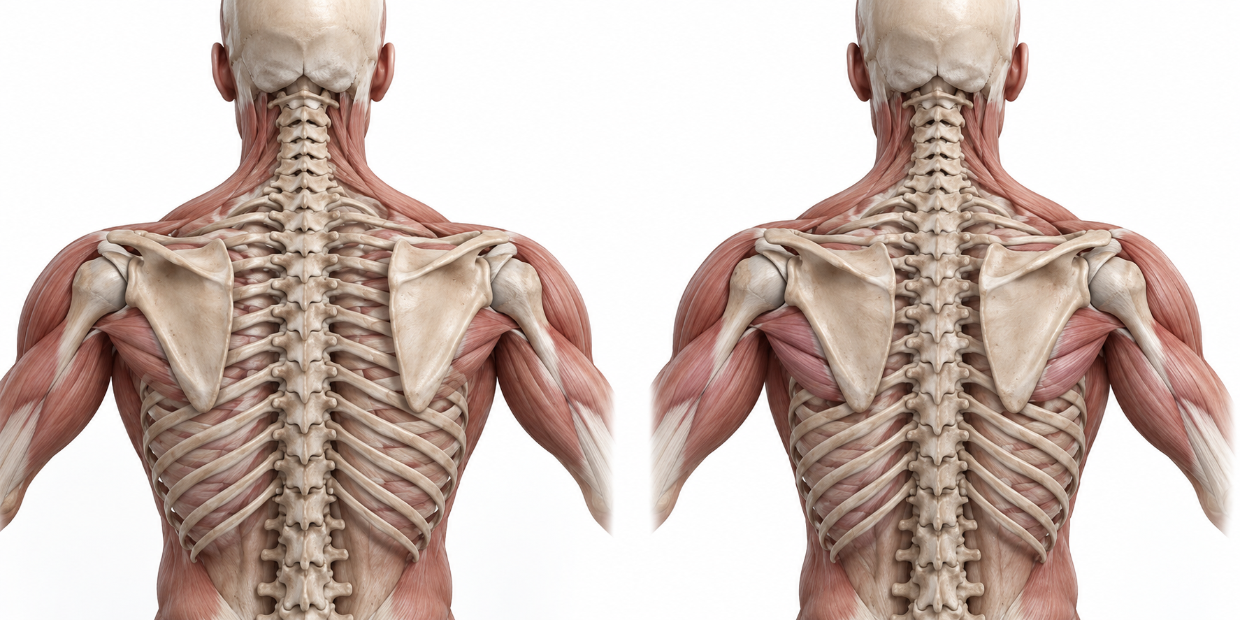
Protraction
Scapulae move around the thorax, away from the spine.
Retraction
Scapulae move closer to the spine over the back of the rib cage.
What the coach asks
Retract, build a base or maintain torso contact.
What the athlete does
Record the setup, repetition phase and visible technique.
What the study resolves
Translation, rotation and tilt need a validated motion record.
Retraction cues can therefore be useful without being read literally as a demand to freeze every scapular degree of freedom for every load and repetition. Research does not justify that universal biological rule.
For a pain-free athlete, the immediate coaching question is usually reproducibility: can the setup be repeated, the unrack controlled and the bar path maintained without an escalating symptom response? For persistent pain, weakness, neurological symptoms, postoperative restrictions or return-to-sport decisions, a cue is not a substitute for clinical assessment.
Layer 05 · Turn “freedom” into a test
How Contact With the Bench May Affect Movement
The posterior torso contacts a surface with a particular width, contour, compliance and friction. Those properties may change edge clearance, contact distribution and the pose available to the athlete. They do not reveal scapular motion by inspection. Article 06 therefore treats pad width as a fit variable, not as a proxy for “mobility.”
Layer 06 · Launch Pad research boundary
The Current Product Evidence Does Not Measure Scapular Motion
The published Launch Pad crossover study measured pectoralis surface EMG, vertical bar displacement, velocity and concentric power in 10 resistance-trained men. It did not measure scapular kinematics.[8] Its complete acute-to-longitudinal evidence treatment belongs to the Athlete-to-Bench Interface; importing bar outcomes as proof of scapular freedom would exceed the data.
Kidwell et al. · 2026 · acute crossover
The published measurements describe the bar and muscle activity
Ten trained men completed five repetitions at 70% 1RM under each condition. Selected bar outcomes are shown below; the article links to the full acute-evidence review.
Vertical bar displacement
SD 7 cm
SD 4 cm
cm · mean with SD · zero-origin scale
Mean bar velocity
SD 0.09 m/s
SD 0.09 m/s
m/s · mean with SD · zero-origin scale
Concentric power
SD 103 W
SD 93 W
W · mean with SD · zero-origin scale
The next decisive study is straightforward in concept and difficult in execution: compare defined surfaces under meaningful loads while synchronizing scapular kinematics, bar forces, contact behavior, performance and symptoms. Until that record exists, the scientifically honest position is neither “scapulae must never move” nor “more movement is always better.” It is: name the motion, locate the phase, identify the method and stop the claim at the measured endpoint.
Next concept · Article 06Bench Pad Width & Shoulder Mechanics: from motion language to fit-envelope testingReferences
- McClure PW, Michener LA, Sennett BJ, Karduna AR. (2001). Direct 3-dimensional measurement of scapular kinematics during dynamic movements in vivo. Journal of Shoulder and Elbow Surgery, 10(3), 269–277. doi:10.1067/mse.2001.112954. PubMed record. ↩
- Ludewig PM, Reynolds JF. (2009). The association of scapular kinematics and glenohumeral joint pathologies. Journal of Orthopaedic & Sports Physical Therapy, 39(2), 90–104. doi:10.2519/jospt.2009.2808. ↩
- Kibler WB, Ludewig PM, McClure PW, Michener LA, Bak K, Sciascia AD. (2013). Clinical implications of scapular dyskinesis in shoulder injury: the 2013 consensus statement from the ‘Scapular Summit’. British Journal of Sports Medicine, 47(14), 877–885. doi:10.1136/bjsports-2013-092425. ↩
- Piepoli A, Miras-Moreno S, Janicijevic D, Martínez-Amat A, García-Ramos A. (2023). Differences in various strength manifestations between the flat and retracted bench press variants: are they affected by subjects’ experience or strength levels? Journal of Strength and Conditioning Research, 37(12), 2339–2345. doi:10.1519/JSC.0000000000004561. ↩
- Noteboom L, Belli I, Hoozemans MJM, Seth A, Veeger HEJ, van der Helm FCT. (2024). Effects of bench press technique variations on musculoskeletal shoulder loads and potential injury risk. Frontiers in Physiology, 15, 1393235. doi:10.3389/fphys.2024.1393235. ↩
- Montenegro S, Izzicupo P, Prestanti I, Serafini S, Fusco A, Sartor F. (2026). Optimizing powerlifting bench press technique using contextual interference via antagonist task selection. Applied Sciences, 16(11), 5511. doi:10.3390/app16115511. ↩
- Tate D. (2008). EFS Bench Manual. elitefts, pp. 8, 12. Practitioner source supplied for this review. Related official elitefts article. ↩
- Kidwell JA, Yamamoto T, Hetherton KJ, Truneh N, Bright JJ, Blatney AE, et al. (2026). Acute effects of thoracic-spinal elevation via a novel bench press pad on sEMG and barbell kinetics in resistance-trained males. International Journal of Exercise Science, 19(1), 1003. doi:10.70252/IJES2026103. ↩